An article in Nature Communications describes how researchers in Italy combined neural manipulations, resting-state fMRI and in vivo electrophysiology to probe how inactivation of a cortical node causally affects brain-wide fMRI coupling in the mouse.
The study was conceived and supervised by Alessandro Gozzi and the electrophysiological recordings were designed and carried out by Federico Rocchi, both of the Functional Neuroimaging Laboratory, Center for Neuroscience and Cognitive systems, Istituto Italiano di Tecnologia, Rovereto, Italy.
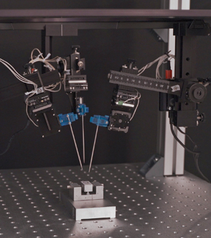
Rocchi used the Multi-Probe Micromanipulator with Virtual Coordinate System (VCS) software to insert three 16-channel Nueronexus silicon neural probes for in vivo electrophysiology recording.
From the paper:
“To measure multi-electrode coherence, three electrodes were inserted in key cortical and subcortical substrates identified as overconnected in our chemo-fMRI mapping in n = 4 hM4Di and n = 5 GFP-expressing mice. A multi-probe micromanipulator (New-Scale Technologies) was used to insert three 16 channels single shank electrode (Neuronexus, USA, interelectrode spacing 1–2.5 mm) in the right prefrontal cortex, centromedial thalamus, and retrosplenial cortex, respectively. Representative electrode locations are reported in Fig. S11, corresponding to the following stereotaxic coordinates… To reduce tissue damage, an insertion rate < 1 μm/min was employed, allowing for a 30-min equilibration every 400 μm traveled… Signals were then recorded into 1 min time bins to cover a 15-min pre-injection baseline and a 40-min post CNO time window.”
Read the article (open access) at https://www.nature.com/articles/s41467-022-28591-3

A Multi-Probe Micromanipulator System at New Scale Technologies, shown with three manipulator arms and steel reference probes.

