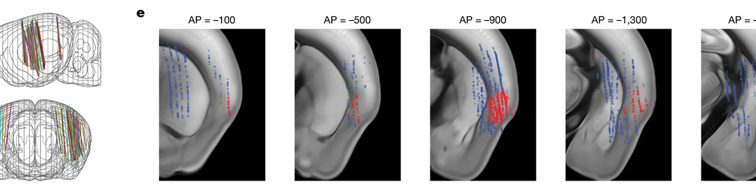
Figure from the recent Deisseroth Lab article in Nature: d, Electrode tracks from n = 5 mice (3 ChRmine and 2 control) over 60 recording sessions co-registered to the common Allen Brain Atlas. e, Locations of recorded single units overlaid onto the Allen Brain Atlas. Red denotes units in the insular cortex. AP, anterior–posterior.
Researchers use MPM Multi-Probe Micromanipulator and four-shank Neuropixels 2.0 probes for in vivo electrophysiology portion of the study
Researchers in the Deisseroth Lab at Stanford University published an article, Cardiogenic control of affective behavourial state, in the 1 March 2023 issue of Nature. The team used the New Scale Technologies Multi-Probe Micromanipulator (MPM) System and Neuropixels 2.0 probes in their work.
From the paper:
Development of a noninvasive optical pacemaker
“In this study, we have developed a method for noninvasive optogenetic control of specific cardiac rhythms during active behaviour… This study shows that cell-type-specific, temporally precise, noninvasive perturbation of organ-scale physiology is possible in fully intact, freely behaving mammals.”
“Our approach… has the potential for broad application to a range of physiological systems throughout the body—opening up numerous opportunities to explore the complex interactions between physiological systems in health, disease and treatment.”
In vivo electrophysiology techniques
“Before recordings, the mice were placed into the pacemaker vests and reliable pacing was confirmed by ECG under brief anaesthesia with isoflurane. Then the mice were head-fixed and allowed to recover.”
“Next, one or two (for simultaneous bilateral recordings) four-shank Neuropixels 2.0 probes mounted on a multi-probe manipulator system (New Scale Technologies) and controlled by SpikeGLX software (Janelia Research Campus) were inserted through the craniotomies at variable angles (0–20°) depending on the recording geometry. Typically the probes were aimed to touch the skull around the insula, which could be inferred from probe bending or changes in local field potential, and then were retracted around 100 µm and allowed to sit in place for at least 15 min before recordings.”
“Recordings were performed along each of the four shanks sequentially while mice received 5 s of optical stimulation (900 bpm (15 Hz)) with inter-trial intervals of at least 15–25 s. Probes were cleaned with trypsin between recording sessions. Spike sorting was performed by Kilosort 2.5 and auxiliary software as previously described.”
Read the open-access article
Hsueh, B., Chen, R., Jo, Y. et al. Cardiogenic control of affective behavioural state. Nature 615, 292–299 (2023). https://doi.org/10.1038/s41586-023-05748-8